Chondrodysplasia
Historical CHD Information
An overview of chondrodysplasia in the Alaskan Malamute and of the Alaskan Malamute Club of America eradication program
Linda M. Dowdy Originally written May 1981 First update October 1999 Second update October 2004 Chondrodysplasia is a genetically-determined skeletal disorder that occurs primarily in the purebred Alaskan Malamute. The purpose of this article is to describe chondrodysplasia and to outline its relation to the veterinary practitioner.


Background
For a number of years Alaskan Malamute breeders in both Canada and the United States have been aware of occasional litters which contained deformed or “dwarfed” puppies. But it was not until the early 1970’s that these puppies were conclusively proven to be the manifestation of a genetic disorder. At first these affected dogs were known as “dwarfs” because of their diminutive size. This term however gave rise to considerable confusion with the dwarfism found in Hereford cattle. So another name, chondrodysplasia, was coined and brought into usage. This disorder, however, does involve far more than simply faulty cartilage maturation. Perhaps when it has been fully understood and defined, then a more comprehensive name will be in order.
Inheritability
Chondrodysplasia is transmitted as a simple autosomal recessive trait. Its mode of inheritance was determined by mating all combinations of unaffected, recessive carrier, and affected animals, until a total of one-hundred-fourteen (114) puppies had been produced. Identification of chondrodysplasia as a simple recessive has led to categorization of the Malamute into three distinct genotypes.
-
Homozygous dominant – clear of the gene (symbol +/+)
-
Heterozygous – recessive carrier of the gene (symbol +/dan)
-
Homozygous recessive – chondrodysplastic (symbol dan/dan)
The term “dan” is taken from a concatenation of the words “dwarfism anemia”. d = dwarfism an = anemia
Phenotype
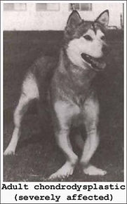
The chondrodysplastic Malamute displays in varying degrees the following phenotypic characteristics.
-
Excessively shortened front limbs with various degrees of bowing and deformity, especially the radius and ulna
-
A topline that slopes from the pelvis down to the withers
Some chondrodysplastic Malamutes display a severe degree of deformity while others display almost no visible characteristics. It is important to note that the recessive carrier genotype displays no physical identifying characteristics, being completely normal in appearance.
Radiographic
Chondrodysplastic puppies, up to twelve weeks of age, are readily identifiable by radiographs of the carpal joint. Although all endochondral bones in the chondrodysplastic animal are affected, the condition is most apparent at the distal end of the ulna. In a normal puppy the distal metaphysis of the ulna appears V-shaped and evenly opaque, consistent with normal ossification and development. In the chondrodysplastic puppy, the distal metaphysis of the ulna is distinctly flattened and irregular, with a blunt widening of the distal diaphysis. There is variable evidence of normal ossification. The physis is increased in width with an irregular metaphyseal margin. After twelve weeks of age the joint begins to change. Some adult chondrodysplastics radiographically display relatively normal ossification. Therefore radiographs should not be assumed as a definitive diagnosis after twelve weeks of age. Radiographic evidence is most consistent between five and twelve weeks of age.
It is important to note that the sloping topline of many chondrodysplastic animals is a manifestation of alteration in conformation of the limbs and is not related to changes in the axial skeleton. The varying degree of topline slope is a direct result of the malformation which develops in the front limbs. The tubular bones at the distal radius and ulna are the most severely affected and growth retardation is excessive. The bones of the hind limbs are much less severely affected and achieve a more normal conformation. Some chondrodysplastic animals may display near normal conformation. Diagnosis of genotype in such cases must then be made by either test-mating the animal or by blood work.
Biochemical Aspects
The chondrodysplastic animal has an associated hemolytic anemia. There are several abnormalities in the red blood cell, including greater size, increased sodium content, and a smaller percentage of solids. The chondrodysplastic also has a trace mineral imbalance, and this eventually results in an excessive accumulation of copper in the liver. Some recessive carriers exhibit the same abnormalities of the red blood cell as the chondrodysplastic, but the overlap with those of clear dogs is excessive.
Blood Profile
As noted earlier, after twelve weeks of age, radiographs no longer provide a positive diagnosis of chondrodysplasia due to changes in the carpal joint. To diagnosis an older animal, it is necessary to utilize a blood profile. Dwarfism affects more than just the front limbs. There is an associated inherited hemolytic anemia that is characterized by morphologically abnormal red cells known as “stomatocytes”. In addition to the stomatocytes, the anemia is characterized by increased red cell size (MCV), decreased mean corpuscular hemoglobin concentration (MCHC), and a normal amount of red cell hemoglobin (MCH). The red cell sodium content is increased while the percent solids is decreased. The following table summarizes some of the work done by Dr. Sheilah Fletch. A second table shows the specific values for three dwarfs. The table of ranges was originally published in the Journal of the American Animal Hospital Association. The left side of the table shows the ranges of three blood parameters (MCHC, MCV, and MCH) for the three phenotypes of the Alaskan Malamute, namely clear, carrier, and chondrodysplastic. The second table shows the same three blood parameters for three known dwarfs (Cameo, Munchkin, and Duke).
Test-Breeding
The AMCA set up a screening and control program to address the problem of chondrodysplasia in the Alaskan Malamute. Initial screening was done by means of pedigree analysis. Using the dog’s pedigree, a statistical analysis was performed based upon the number of ancestors that were either known carriers or closely related to known carriers. The analysis yielded a number expressed as a percentage. This number represented the dog’s chances of being a recessive carrier of the gene, based upon the configuration of ancestors. If the number exceeded 6.25%, the dog was considered to be in the “suspect” or “potential” category. The value of 6.25% was selected because it represented one known carrier as a great-great-great-grandparent and also corresponds roughly to a 95% confidence level (04.37%). If the pedigree analysis indicated the animal fell into the “suspect” category, two recommendations were made to the owner.
-
Withhold the animal from all breeding and preferably have it neutered
-
Withhold the animal from all breeding until it could be test-mated with a chondrodysplastic or known recessive carrier.
Another method of testing, based on the blood profile of the animal, saw limited utilization. It was considered superior to pedigree analysis but not as good as test-breeding.
Genetics
Let the symbols for the various genotypes be as follows.
-
dan/dan = chondrodysplastic animal
-
+/dan = recessive carrier
-
+/+ = clear animal
-
+/? = unknown genotype animal
In test-mating with a chondrodysplastic animal, then: (+/?) X (dan/dan) => 1/2(+/dan) + 1/2(?/dan)
If all the test puppies in the litter are radiographically normal, then ? = +, and the genotype of the unknown is statistically identified as being clear. If however one or more chondrodysplastic puppies are detected, then ? = dan, and the suspect animal is conclusively proven to be a recessive carrier. Such an animal should be removed from all further breeding, unless it is the control dog in the test-mating of another suspect animal.
Radiographing Test Litters
Test litters are normally radiographed at four to six weeks of age. A high-detail A/P projection of the distal radius and ulna of one leg is needed of each puppy in the litter. Several puppies may be included on one sheet of film, thus reducing costs for the owner. Utilization of lead or leaded vinyl/rubber sheets as film blockers (or close collimation) will avoid radiation fogging on multiple exposures. The film should be clearly marked with the owner’s name, name of sire, name of dam, number of puppies in the litter, and date of birth of the puppies. Any dead puppies that have been preserved by freezing should be radiographed at this time. These are frequently inconclusive due to age, however, and it should be understood they may not be included in statistical evaluation unless chondrodysplasia is clearly evident. [reference 8] Radiographs must be evaluated by a professional who is familiar with chondrodysplasia, and the following information should be included.
-
Owner’s name and address
-
Sire’s name
-
Dam’s name
-
Status of control animal (chondrodysplastic or recessive carrier)
-
Number of puppies in litter and date of birth
-
Number of puppies x-rayed and date of x-rays
Disposition of Test-Litter Puppies
Test-litter puppies, even though normal in appearance, must not be allowed to slip back into open breeding. Where one parent is a chondrodysplastic, all normal puppies in the test litter are recessive carriers of the gene. The AMCA approved of the following methods for handling test puppies.
-
Donation to institution working on problem of chondrodysplasia
-
Euthanasia by humane means
-
Early sterilization and subsequent placement in pet homes
Early sterilization encompasses tubal ligation on puppy bitches and vasectomy on puppy dogs. It is normally done between five and eight weeks of age. The AMCA produced a brochure on the early sterilization process. The only exceptions to the stated methods of disposition of test puppies would be the holding of chondrodysplastics, or possibly carriers, to be used as control animals in further test-breeding.
Blood-Testing
To a very limited extent, hematological testing was utilized as a third method of screening dogs. The process did not prove to be a valuable weapon in AMCA’s arsenal however. The necessity of baselining each laboratory’s instruments and procedures against a control group of known genotype dogs in order to establish normal values ultimately made the process infeasible. In blood testing, eleven different parameters were measured from a sample of blood. These parameters were then submitted to a statistical computer program that enhanced differences and weighted all values in order to determine the most likely genotype for that particular blood sample. Misclassification rate on the control group of fifty dogs was approximately 6% in either direction. Perhaps in the final analysis, the immensely valuable contribution of the blood test will be recognized as having conclusively demonstrated that the heterozygous animal has measurable differences from the clear animal.
Tubal Ligation and Vasectomy
This brochure, produced by the Alaskan Malamute Club of America, Inc., describes a method of rendering very young puppies incapable of reproduction. The procedures described were developed by AMCA member John Schmidt DVM and fellow veterinarian Dan F. Rice DVM. They were originally developed for the safe handling of puppies produced by test-breedings for chondrodysplasia, a genetic disorder that affects Alaskan Malamutes. For many years breeders have wanted a method of birth control that would render very young puppies incapable of breeding so that they could be sold as pets and not become a part of the breeding population. It is important to breeders to have these procedures completed before a pup is seven to eight weeks old as many prefer to make placements at that age. Such results may be obtained by salpingectomy (tubal ligation) in the bitch puppy when she is as young as three weeks old and by vasectomy in the male puppy when he is about 5 1/2 weeks old. These procedures are acceptable by the Chondrodysplasia Committee for handling puppies from test litters. Puppies should be examined carefully prior to surgery. They may be anesthetized and prepared for surgery by the standard methods used in any veterinary practice. In the female a small incision is made into the abdominal cavity caudal to the umbilicus and the ovary is elevated through the incision. The oviduct is easily seen in the mesovarium (see figure 1). It is grasped with forceps; a small section is removed and the cut ends are cauterized. The same procedure is repeated on the opposite side. Both ovaries are returned to the abdominal cavity and the abdominal incision is closed by standard methods. Please note that only a small portion of the oviduct is removed. The uterus and ovaries are left intact. Vasectomy in the male may be done as soon as the testicles are descended to the scrotum, usually when the puppy is about five weeks old. A single small midline incision is made cranial to the scrotum and a spermatic cord is elevated through the opening. A small incision is then made in the common vaginal tunic of the spermatic cord. The ductus deferens is located in a medial fold of the cord along with a small artery and vein (see figure 2 and 3). The ductus deferens is separated from the artery and vein; a small section is removed and the cut ends are cauterized. The common vaginal tunic is closed around the spermatic cord but usually no sutures are required to close this incision. The procedure is then repeated on the opposite cord. Fascia and skin are closed by standard methods. Again please note that only a small portion of the ductus deferns is removed. The testicles remain intact. Prospective owners of the pet puppies should be fully informed of the procedures that have been done. A bitch will still come into estrus and show all the signs of a normal heat. She will stand for a male but cannot conceive. It is suggested that bitches be confined when in heat and should be spayed at the usual time recommended by the veterinarian. This will prevent the undesirable manifestations of estrus and eliminate the possibility of later reproductive tract problems.
Figure 1 and 2 are from: “Anatomy of the Dog”:Miller, Christiensen & Evans. W.B. Saunders Co., Philadelphia. Figure 3: “Guide to the Dissection of the Dog”: Miller. Lithographed by Edwards Bros. Inc., Ann Arbor, Mich. Cornell Univeristy, Ithaca, New York. 1952
REFERENCES
1. Subden RE, Fletch SM, Smart ME, Brown RG “Genetics of the Alaskan Malamute Chondrodysplasia Syndrome” Journal of Heredity 63:149,1972 2. Sande RD, Alexander JE, Padgett GA “Dwarfism in the Alaskan Malamute: Its Radiographic Pathogenesis” Journal of the American Veterinary Radiology Society, 15, 1974 Sande RD, Pennock PW, Burt JK Personal correspondence, 1978 3. Sande RD Personal correspondence, 1978 4. Pinkerton PH, Fletch SM “Inherited Haemolytic Anaemia With Dwarfism in the Dog” Blood, 40:963,1972 Pinkerton PH, Fletch SM, Bruckner PJ,Miller DR “Hereditary Somtatocytosis With Hemolytic Anemia in the Dog” Blood, 44:557,1974 5. Brown RG, Hoag GN, Subden RE, Smart ME “Alaskan Malamute Chondrodysplasia I – V” Growth, Vol 41, 1977 6. Fletch SM, Pinkerton PH, Bruckner PJ “The Alaskan Malamute Chondrodysplasia (Dwarfism – Anemia) Syndrome in Review” Journal of theAmericanAnimalHospital Association, Vol 2 No. 3, 353-361,1975 7. Bourns TKR, Dodd HM, Dowdy LM, Lucus AJ, Pearson DS “The Master Plan for Control and Elimination of the Condition Known as Dwarfism” Alaskan Malamute Club ofAmerica Newsletter, November 1971 8. Sande RD, Pennock PW, Burt JK Personal correspondence, 1978 9. Schmidt JE “Tubal Ligation and Vasectomy” Brochure available from the Alaskan Malamute Club of America Online at Tubal Ligation and Vasectomy” Rice DF, Dewell CG “Sterilization of Nursing Puppies” Modern Veterinary Practice (Clinical Reports), Vol. 57, No. 10, Oct. 1976 10. Fletch, SM Personal correspondence, 1976-1977
PHOTO CREDITS
“Nori”, severely affected chondrodysplastic Photo by Bill Francis Radiographs – Radiographs of chondrodysplastic and phenotypic normal courtesy of Dept. of Radiology, Ohio State University
CONTRIBUTORS AND ADVISORS
My thanks goes to the following people who contributed their time and knowledge. Without their help and advise, this article would not have been possible.
-
Beckman, JM
-
Bourns, TKR
-
Burt, JK
-
Cox, MD
-
Fletch, SM</sp an>
-
Pearson, DS
-
Pennock, PW
-
Sande, RD
-
Schmidt, JE
-
Smart, ME

The status of obtaining a genetic test for CHD
July 8, 2012: The researchers have identified approximately 30 variants of interest in our samples, and are running these through further analysis. Additionally, they will be doing a SNP-chip genotyping study which will hopefully increase our chances to find the causative mutation. Additional new samples from carriers and an affected dog are being sent to the lab, which will help Dr. Lohi’s group confirm any findings they may uncover. We should know more in the coming few months. We have informed them that if any funds are needed to accelerate the research, we will do what is needed to provide these through the Canine Health Foundation grant process using our Donor Advised Fund. January, 2012: At the beginning of 2011, we were told that the researcher at MSU who has been working on this project for years, Dr. Pat Venta, was willing to collaborate with another researcher who had more advanced technology in an effort to find this gene.
Unfortunately, several months later we were informed that he had decided against this collaboration and wanted to pursue a grant on his own to do the research. At this time no grant has been pursued. Because finding the gene for dwarfism is a priority, we are exploring all other options for this research. Dr. Hannes Lohi at the University of Helsinki discovered the gene for dwarfism in Norwegian Elkhounds, but samples from our dwarfs have found that this is not the same gene. However, his research team has taken on finding this gene in our breed-at no cost. We have gathered samples from all currently living dwarfs and dwarf carriers and have shipped the majority of these to their facility in Helsinki. These samples have been run through a whole exome scan platform, which is the most advanced genetic analysis available. They hope to have the results from this in a few months, at which time they will look for abnormalities which may represent the gene for the disease. How long this will take depends completely upon what the genetic results look like, but we feel confident that this team is doing everything that can be done on this project as quickly as is possible. They have been extremely responsive to any questions we have, and have been very proactive in asking us questions as well as getting any samples into their lab. If any new dwarfs are identified, we desperately need DNA from these dogs for our research study. If you have any samples to contribute or questions, please feel free to contact: Sandi Shrager AMCA Health Committee Chair sandis@u.washington.edu 206-919-8103206-919-8103 Kaisa Kyostila kaisa.kyostila@helsinki.fi Ranja Eklund/Lohi Laboratory Biomedicum Helsinki, room B320 Haartmaninkatu 8 00290 Helsinki Finland Below is a table with descriptions of the type abbreviations used in the database:
Chondrodysplasia
Chondrodysplasia is a genetic disorder in the Alaskan Malamute which manifests itself in puppies born with deformities, eventually evident in the abnormal shape and length of their limbs. Chondrodysplasia is present in adult carriers as an auto-somal or simple recessive gene. Therefore, both sire and dam must carry this gene in order to produce an affected (chondrodysplastic) puppy. As Malamute owners and prospective owners, we have an opportunity to control the proliferation of Chondrodysplasia in the Alaskan Malamute, and with such control, there is hope of its total elimination in our breed.
History and Explanation
For a number of years, Alaskan Malamute breeders in both the United States and Canada were aware of occasional litters that contained deformed or “dwarf puppies”, produced by parents who showed no physical evidence of the condition. It was not until the early 1970’s that these puppies were conclusively proven to be the manifestation of a genetic disorder. At first, these affected dogs were known as “dwarves” because of their diminutive size. This term gave rise to considerable confusion. Veterinarians associated it with the dwarfism found in Hereford cattle, while owners associated it with any small Malamute. Since neither is correct, another name, Chondrodysplasia(meaning “faulty cartilage”), was coined and brought into usage. Chondrodysplasia in the Malamute was originally diagnosed as a form of rickets. Upon closer examination at various veterinary schools, it was determined that this diagnosis was incorrect. While it isn’t known exactly what this crippling problem is, it has definitely been proven to be genetic, or inherited. A more technical description of this gene is “auto-somal,” or “simple recessive.” This simply means that the sire and dam must both carry this gene in order to produce an affected chondrodysplastic puppy. In very young puppies, under six weeks of age, the deformity is often very difficult, if not impossible, to detect without x-rays, even to the practiced eye. But as the puppies grow older, the deformity becomes more evident in the shape and length of the front legs. However, not all chondrodysplastics are severely affected. In some adults, the front legs may appear “almost” normal. It is not the chondrodysplastics themselves that are the major problem. The greatest concern is the use in breeding of the completely normal appearing dogs that possess or “carry” this gene. (A good parallel of this among humans is two brown-eyes parents producing a blue-eyed baby. In spite of their brown eyes, the parents both “carry” the gene for the blue eye.) While the breeding of two carriers can produce a chondrodysplastic, a litter from a breeding between a carrier and a “non carrier” (or clear) will contain only normal appearing dogs. Nevertheless, an undetermined number of puppies will themselves be carriers, having inherited the gene from their carrier parent. The continued whelping of such litters increase the number of carriers “at large” in the total Malamute population.
Chondrodysplasia Certification Registry
CHD Certification is available to dogs with one certified ancestor between the applicant dog and each uncertified ancestor. If proof can be offered that an uncertified ancestor is a full sibling to a certified dog, the applicant’s pedigree will be evaluated. It should be noted that certification can be revoked when information is verified that a dog was mis-classified through subsequent breedings and/or test breedings. Sometimes the pedigree reveals the dog to be non-certifiable with regards to Chondrodysplasia. In such cases, the AMCA recommends these dogs not be bred. The owner may feel the dog to be a great asset to a breeding program, or the dog may already have offspring who are themselves now affected by the un-certifiable status of the parent. In such circumstances, the owner may choose to apply for a further screening process known as “Test Breeding”. The certificate is not proof that a dog is clear
(i.e. not a carrier), but only that the probability that it is is calculated to be below 6.25%. AMCA is supporting genetic research programs with the intent of finding a DNA marker allowing the proof that a dog is either clear of this genetic disorder or is not. Whenever this test is available, then there will no longer be a need for test breeding to track this condition.
Test Breeding
The committee will assist the owner in this choice. At present, test breeding a suspect Malamute to a Chondrodysplastic, or to a known carrier, is the only means to be reasonably certain the dog is not a carrier of the gene. Should the owner decide to enter the suspect dog into a test breeding program, a contract specifying certain criteria is entered into with the AMCA Chondrodysplasia Certification Committee. The Committee Chairman and Area Representatives are available for consultation regarding the procedures for test breeding. Once thought to occur only in Malamute, Chondrodysplasia has now been detected in other breeds and the AMCA program has become a model for other breed clubs. Your efforts are needed if it is to continue to fulfill its vital leadership role. We hope you will join us in the fight to control this genetic disorder. It is essential for the prospective malamute purchaser of potential breeding stock to realize the importance of these certificates. By purchasing from AMCA Certified stock, the buyer not only is helping the breed cull out Chondrodysplasia, but is also relieving himself of the responsibility of test breeding.
---
The Status of Obtaining a Genetic Test for Chondrodysplasia
Dr. Lohi has released the following Report to the Canine Health Foundation on the current progress of our research project.
Report to Grant Sponsor from Investigator:
“In the Alaskan Malamute chondrodysplasia study, we have been using two complementary research methods to discover the causative mutation: exome sequencing and DNA-marker based genome-wide association mapping. Thus far we have analyzed genome-wide data from ten affected dogs and up to 87 healthy control dogs.
Part of the control cohort was obtained from a previous cataract study. The Alaskan Malamute chondrodysplasia is thought to have an autosomal recessive mode of inheritance and we have performed our data analyses under this model. We have now identified a chromosomal region that shows association to the disease. This region was identified by using several different genome-wide analysis approaches, which included both basic case-control association and family-based methods.
The finding is still tentative because our sample cohort has some issues concerning unwanted population structure. The affected dogs form a separate cluster from the control samples, which means that they come from a separate line of dogs than the control dogs. This population structure can have an effect to the results by introducing false positive findings that are caused by the population difference and not by the difference in affection status.
To correct this problem, we have recently genotyped a new sample set of seven affected and seven control dogs but unfortunately half of these sample failed because of their poor quality. We have previously performed exome sequencing to one carrier dog and two affected dogs, and now we are in theprocess of sequencing four additional affected and two additional carrier dogs by using an updated exome sequencing platform.
The main obstacle in this study has been the availability and quality of samples. Many samples of affected dogs have been received as old DNA samples that have had both low DNA-quality and quantity. Furthermore, control dogs that are closely related to the affected dogs have been difficult to obtain, and this has caused the problems in population structure.
Consequently, any new samples from affected dogs and their relatives would be greatly appreciated. However, people within the Alaskan Malamute breed have been extremely supportive of the research project, and we wish to warmly thank all who have contributed to the study and helped us in obtaining samples.”
Dr. Lohi’s research group has just received a set of samples from Dr. Venta’s original research project. This represents a number of additional affected and carrier dogs and should greatly facilitate the research project and progress.
Dr. Lohi has applied for and received an Acorn Grant through the American Kennel Club’s Canine Health Foundation to help facilitate the research project. This grant has been fully funded by the Alaskan Malamute Research Foundation.
http://www.akcchf.org/research/funded-research/1901.html
The researchers have checked 28 variants from the exome-data in a bigger sample cohort but unfortunately none of these variants showed perfect segregation with the disease. They are currently performing the lab work to check another set of 27 variants (ioinformatician re-analyzed the exome-data with another program and with some new interesting variants that they are looking through).
The researchers have identified approximately 30 variants of interest in our samples, and are running these through further analysis. Additionally, they will be doing a SNP-chip genotyping study which will hopefully increase our chances to find the causative mutation.
Additional new samples from carriers and an affected dog are being sent to the lab, which will help Dr. Lohi’s group confirm any findings they may uncover. We should know more in the coming few months. We have informed them that if any funds are needed to accelerate the research, we will do what is needed to provide these through the Canine Health Foundation grant process using our Donor Advised Fund.
At the beginning of 2011, we were told that the researcher at MSU who has been working on this project for years, Dr. Pat Venta, was willing to collaborate with another researcher who had more advanced technology in an effort to find this gene. Unfortunately, several months later we were informed that he had decided against this collaboration and wanted to pursue a grant on his own to do the research.
At this time no grant has been pursued. Because finding the gene for dwarfism is a priority, we are exploring all other options for this research. Dr. Hannes Lohi at the University of Helsinki discovered the gene for dwarfism in Norwegian Elkhounds, but samples from our dwarfs have found that this is not the same gene. However, his research team has taken on finding this gene in our breed–at no cost.We have gathered samples from all currently living dwarfs and dwarf carriers and have shipped the majority of these to their facility in Helsinki.
These samples have been run through a whole exome scan platform, which is the most advanced genetic analysis available. They hope to have the results from this in a few months, at which time they will look for abnormalities which may represent the gene for the disease. How long this will take depends completely upon what the genetic results look like, but we feel confident that this team is doing everything that can be done on this project as quickly as is possible. They have been extremely responsive to any questions we have, and have been very proactive in asking us questions as well as getting any samples into their lab.


